产品号 #05715_C
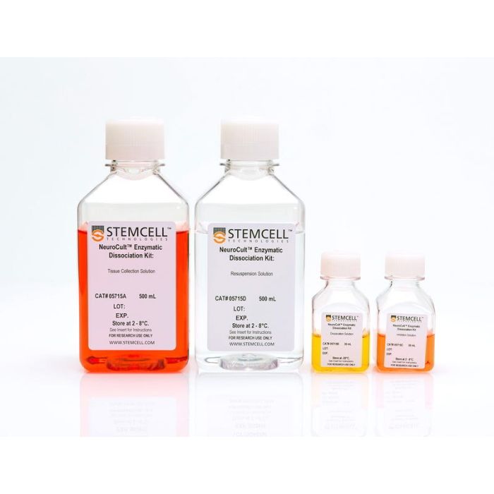
成年小鼠和大鼠CNS组织酶解试剂盒
NeuroCult™成年中枢神经系统(CNS)组织酶解试剂盒(小鼠和大鼠)推荐用于成年小鼠和大鼠(CNS)组织的酶消化和解离。NeuroCult™酶解试剂盒已经过优化,整个过程快速、可重复,并可获得高活率和高数量的细胞。由此获得的单细胞悬液可立即用于下游应用。
亚型
酶法相关(或酶解类产品
细胞类型
神经干/祖细胞
种属
小鼠,大鼠
品牌
NeuroCult
研究领域
神经科学,干细胞生物学
Find supporting information and directions for use in the Product Information Sheet or explore additional protocols below.
This product is designed for use in the following research area(s) as part of the highlighted workflow stage(s). Explore these workflows to learn more about the other products we offer to support each research area.
| Species | Mouse, Rat |
|---|
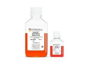
小鼠和大鼠神经干细胞及祖细胞扩增培养基套装
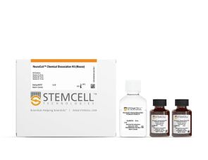
小鼠神经球化学解离试剂盒
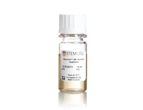
无血清神经添加物(50X)
提升神经元功能的无血清基础培养基