EasySep™ Release人PE正选试剂盒

采用可解离磁珠技术对PE偶联抗体标记的人的细胞进行免疫磁珠正选

产品号 #(选择产品)

产品号 #17654_C

对PE偶联抗体标记的无颗粒人细胞进行免疫磁珠阳性分选

产品优势

  • 只需不到40分钟,从人或小鼠组织中分离出PE抗体标记的高纯度细胞
  • 无需洗涤去除EasySep™ Releasable RapidSpheres™可解离磁珠

产品组分包括

  • EasySep™ Release人PE正选试剂盒(产品号 #17654)
    • EasySep™ Release PE正选抗体混合物,1 mL
    • EasySep™ Releasable RapidSpheres™ 50201磁珠,1 mL
    • EasySep™Release缓冲液(浓缩),3 x 1 mL
    • 抗人CD32阻断剂,1 mL
New look, same high quality and support! You may notice that your instrument or reagent packaging looks slightly different from images displayed on the website, or from previous orders. We are updating our look but rest assured, the products themselves and how you should use them have not changed. Learn more

总览

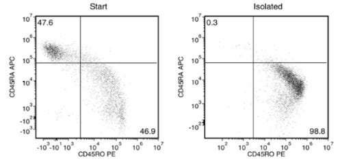
使用 EasySep™ Release人PE正选试剂盒,可通过免疫磁性正选,轻松从新鲜或冻存的人外周血单个核细胞(PBMCs)及经洗涤的白细胞单采样本中分离出高纯度、且不含磁珠的藻红蛋白PE偶联抗体标记人的细胞。EasySep™技术结合单克隆抗体的特异性和无柱磁分选系统的简便性,已在发表的研究中广泛应用超过20年。

在该EasySep™阳性分选流程中,首先用能识别PE和磁珠(称为EasySep™可解离磁珠)的抗体复合物标记目标细胞。与传统磁珠结合目标细胞不同,这些磁珠具有可解离的特性。经EasySep™磁极分选后,使用解离试剂可将结合的磁珠从EasySep™分选的PE抗体标记细胞上移除,非目标细胞则被定向去除。最终分选获得的细胞组分含有高纯度PE阳性细胞,可立即用于流式细胞术、细胞培养或DNA/RNA提取等下游应用。使用该EasySep™试剂盒分选之后,细胞表面仍结合有抗体复合物,并可能与Brilliant Violet™偶联的抗体、聚乙二醇修饰的蛋白质或其他化学相关配体相互作用。

本产品替代EasySep™ Human PE正选试剂盒(货号#18551),可提供高纯度无磁珠标记的细胞。

了解更多关于免疫磁珠EasySep™技术的工作原理。探索更多为您的实验流程优化的产品,包括培养基、添加剂、抗体等配套试剂。

磁体兼容性
• EasySep™ Magnet (Catalog #18000) • “The Big Easy” EasySep™ Magnet (Catalog #18001) • EasyPlate™ Magnet (Catalog #18102) • EasyEights™ Magnet (Catalog #18103)
 
亚型
细胞分选试剂盒
 
细胞类型
B 细胞,树突状细胞(DCs),粒细胞及其亚群,造血干/祖细胞,巨噬细胞,骨髓基质细胞,间充质干/祖细胞,单核细胞,单个核细胞,髓系细胞,NK 细胞,其它细胞系,血浆,T 细胞
 
种属

 
样本来源
Leukapheresis,其它细胞系,PBMC
 
筛选方法
Positive
 
应用
细胞分选
 
品牌
EasySep
 
研究领域
免疫
 

Data Figures

Typical EasySep™ Release Human PE Positive Selection Profile

Figure 1. Typical EasySep™ Release Human PE Positive Selection Profile

Starting with fresh PBMCs, the purities of the start and final isolated fractions are 46.9% and 98.8%, respectively, using a PE-conjugated anti-human CD45RO antibody and EasySep™ Release Human Positive Selection Kit.

Protocols and Documentation

Find supporting information and directions for use in the Product Information Sheet or explore additional protocols below.

Document Type
Product Name
Catalog #
Lot #
Language
Catalog #
17654
Lot #
1000159295 or higher
Language
English
Document Type
Safety Data Sheet 1
Catalog #
17654
Lot #
All
Language
English
Document Type
Safety Data Sheet 2
Catalog #
17654
Lot #
All
Language
English
Document Type
Safety Data Sheet 3
Catalog #
17654
Lot #
All
Language
English
Document Type
Safety Data Sheet 4
Catalog #
17654
Lot #
All
Language
English

Resources and Publications

Educational Materials (11)

Publications (4)

Tumor-Infiltrating PD-L1+ Neutrophils Induced by GM-CSF Suppress T Cell Function in Laryngeal Squamous Cell Carcinoma and Predict Unfavorable Prognosis. D. Tang et al. Journal of inflammation research 2022

Abstract

PURPOSE Chronic inflammation contributes to tumor initiation, progression, and immune escape. Neutrophils are the major component of inflammatory response and participate in the tumorigenesis process. However, compared to other immune cells in the tumor microenvironment of laryngeal squamous cell carcinoma (LSCC), neutrophils, especially the tumor-associated neutrophils (TANs), have not yet been comprehensively explored. The mechanism for regulating the crosstalk between TANs and tumor cells still remains unclear. MATERIALS AND METHODS The distribution profiles and phenotypic features of neutrophils and other inflammatory immune cell populations from a large LSCC patient cohort were systemically analyzed. Co-culturing of peripheral blood associated neutrophils (PANs) and TANs with PBMCs was performed, and the immunosuppression effect on T-cells was examined. RESULTS LSCC microenvironment is highly inflammatory with remarkable TANs infiltration, which is often associated with unfavorable prognosis and advanced clinical stage. We find that TANs in LSCC display morphologically immature and lower apoptosis, exhibit distinctively immunosuppressive phenotype of high PD-L1, and suppress CD8+ T lymphocytes proliferation and activation. We subsequently discover that PD-L1+TANs induced by LSCC-derived GM-CSF potently impair CD8+ T-cells proliferation and cytokines production function, which are partially blocked by a PD-L1-neutralizing antibody. Clinical data further support GM-CSF as an unfavorable prognostic biomarker and reveal a potential association with inflammatory immune cell infiltration, in particular neutrophils. CONCLUSION Tumor-infiltrating PD-L1+ neutrophils induced by LSCC-derived GM-CSF suppress T cell proliferation and activation in the inflammatory microenvironment of LSCC and predict unfavorable prognosis. These TANs cripple antitumor T cell immunity and promote tumor progression. Our findings provide a basis for targeting PD-L1+TANs or GM-CSF as a new immunotherapeutic strategy for LSCC.
STING differentially regulates experimental GVHD mediated by CD8 versus CD4 T cell subsets. C. S. Bader et al. Science translational medicine 2020 jul

Abstract

The stimulator of interferon genes (STING) pathway has been proposed as a key regulator of gastrointestinal homeostasis and inflammatory responses. Although STING reportedly protects against gut barrier damage and graft-versus-host disease (GVHD) after major histocompatibility complex (MHC)-mismatched allogeneic hematopoietic stem cell transplantation (aHSCT), its effect in clinically relevant MHC-matched aHSCT is unknown. Studies here demonstrate that STING signaling in nonhematopoietic cells promoted MHC-matched aHSCT-induced GVHD and that STING agonists increased type I interferon and MHC I expression in nonhematopoietic mouse intestinal organoid cultures. Moreover, mice expressing a human STING allele containing three single-nucleotide polymorphisms associated with decreased STING activity also developed reduced MHC-matched GVHD, demonstrating STING's potential clinical importance. STING-/- recipients experienced reduced GVHD with transplant of purified donor CD8+ T cells in both MHC-matched and MHC-mismatched models, reconciling the seemingly disparate results. Further examination revealed that STING deficiency reduced the activation of donor CD8+ T cells early after transplant and promoted recipient MHC class II+ antigen-presenting cell (APC) survival. Therefore, APC persistence in STING pathway absence may account for the increased GVHD mediated by CD4+ T cells in completely mismatched recipients. In total, our findings have important implications for regulating clinical GVHD by targeting STING early after aHSCT and demonstrate that an innate immune pathway has opposing effects on the outcome of aHSCT, depending on the donor/recipient MHC disparity.
Rheumatoid Arthritis Patients, Both Newly Diagnosed and Methotrexate Treated, Show More DNA Methylation Differences in CD4+ Memory Than in CD4+ Na\ive T Cells." K. Guderud et al. Frontiers in immunology 2020

Abstract

Background: Differences in DNA methylation have been reported in B and T lymphocyte populations, including CD4+ T cells, isolated from rheumatoid arthritis (RA) patients when compared to healthy controls. CD4+ T cells are a heterogeneous cell type with subpopulations displaying distinct DNA methylation patterns. In this study, we investigated DNA methylation using reduced representation bisulfite sequencing in two CD4+ T cell populations (CD4+ memory and na{\{i}}ve cells) in three groups: newly diagnosed disease modifying antirheumatic drugs (DMARD) na{\"{i}}ve RA patients (N = 11) methotrexate (MTX) treated RA patients (N = 18) and healthy controls (N = 9) matched for age gender and smoking status. Results: Analyses of these data revealed significantly more differentially methylated positions (DMPs) in CD4+ memory than in CD4+ na{\""{i}}ve T cells (904 vs. 19 DMPs) in RA patients compared to controls. The majority of DMPs (72{\%}) identified in newly diagnosed and DMARD na{\""{i}}ve RA patients with active disease showed increased DNA methylation (39 DMPs) whereas most DMPs (80{\%}) identified in the MTX treated RA patients in remission displayed decreased DNA methylation (694 DMPs). Interestingly we also found that about one third of the 101 known RA risk loci overlapped (±500 kb) with the DMPs. Notably introns of the UBASH3A gene harbor both the lead RA risk SNP and two DMPs in CD4+ memory T cells. Conclusion: Our results suggest that RA associated DNA methylation differences vary between the two T cell subsets but are also influenced by RA characteristics such as disease activity disease duration and/or MTX treatment."""

更多信息

更多信息
Species Human
Magnet Compatibility • EasySep™ Magnet (Catalog #18000) • “The Big Easy” EasySep™ Magnet (Catalog #18001) • EasyPlate™ Magnet (Catalog #18102) • EasyEights™ Magnet (Catalog #18103)
Sample Source Leukapheresis, Other, PBMC
Selection Method Positive
PRODUCTS ARE FOR RESEARCH USE ONLY AND NOT INTENDED FOR HUMAN OR ANIMAL DIAGNOSTIC OR THERAPEUTIC USES UNLESS OTHERWISE STATED. FOR ADDITIONAL INFORMATION ON QUALITY AT STEMCELL, REFER TO WWW.STEMCELL.COM/COMPLIANCE.
版权 © 2025 STEMCELL Technologies 技术有限公司。保留所有权利。