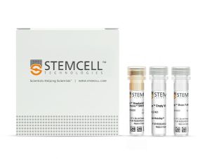
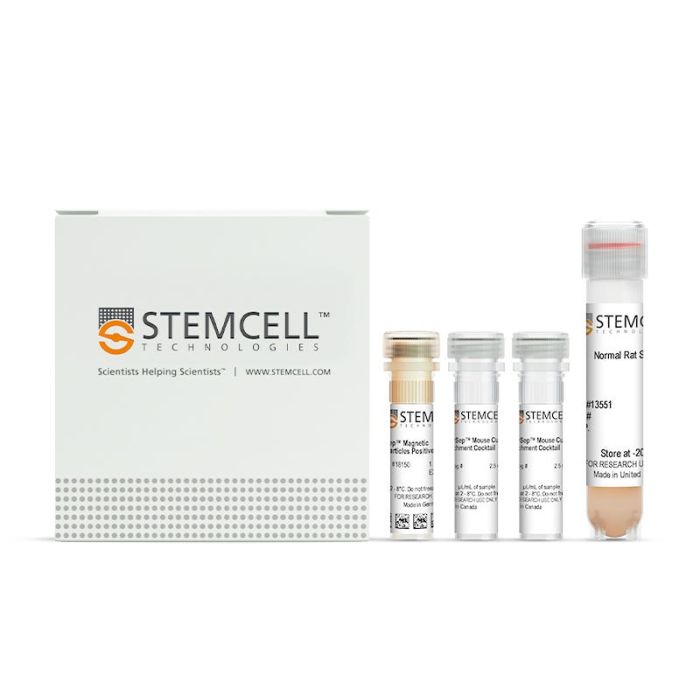
EasySep™ Mouse Custom Enrichment Kit

Immunomagnetic positive selection kit

产品优势


  • Fast and easy-to-use

  • Up to 98% purity

  • No columns required

  • Isolated cells are not fluorochrome-labeled

  • The EasySep™ Mouse CD90.2 Positive Selection Kit II is designed to isolate CD90.2+ (Thy1.2+) cells from single-cell suspensions of splenocytes or other tissues by positive selection. Desired cells are targeted with antibody complexes recognizing CD90.2 and dextran-coated magnetic particles. Labeled cells are separated using an EasySep™ magnet without the use of columns. Cells of interest remain in the tube while unwanted cells are poured off.

    This product replaces the EasySep™ Mouse CD90.2 Positive Selection Kit (Catalog #18751) for even faster cell isolations and does not result in the labeling of isolated cells with PE.

    Data Figures

    Typical EasySep™ CD902 Positive Selection Profile

    Figure 1. Typical EasySep™ CD90.2 Positive Selection II Profile

    Starting with mouse splenocytes, the CD90.2+ cell content of the isolated fraction is typically 95.8 ± 1.5% (mean ± SD using the purple EasySep™ Magnet).

    Protocols and Documentation

    Find supporting information and directions for use in the Product Information Sheet or explore additional protocols below.

    Document Type
    Product Name
    Catalog #
    Lot #
    Language
    Catalog #
    18951RF
    Lot #
    All
    Language
    English
    Catalog #
    18951
    Lot #
    All
    Language
    English
    Document Type
    Safety Data Sheet 1
    Catalog #
    18951RF
    Lot #
    All
    Language
    English
    Document Type
    Safety Data Sheet 2
    Catalog #
    18951RF
    Lot #
    All
    Language
    English
    Document Type
    Safety Data Sheet 3
    Catalog #
    18951RF
    Lot #
    All
    Language
    English
    Document Type
    Safety Data Sheet 4
    Catalog #
    18951RF
    Lot #
    All
    Language
    English
    Document Type
    Safety Data Sheet 1
    Catalog #
    18951
    Lot #
    All
    Language
    English
    Document Type
    Safety Data Sheet 2
    Catalog #
    18951
    Lot #
    All
    Language
    English
    Document Type
    Safety Data Sheet 3
    Catalog #
    18951
    Lot #
    All
    Language
    English

    Applications

    This product is designed for use in the following research area(s) as part of the highlighted workflow stage(s). Explore these workflows to learn more about the other products we offer to support each research area.

    Resources and Publications

    Educational Materials (9)

    Publications (2)

    Mannan-Binding Lectin Promotes Murine Graft-versus-Host Disease by Amplifying Lipopolysaccharide-Initiated Inflammation. D. Heja et al. Transplantation and cellular therapy 2022 aug

    Abstract

    Conditioning regimens used for hematopoietic stem cell transplantation (HCT) can escalate the severity of acute T cell-mediated graft-versus-host disease (GVHD) by disrupting gastrointestinal integrity and initiating lipopolysaccharide (LPS)-dependent innate immune cell activation. Activation of the complement cascade has been associated with murine GVHD, and previous work has shown that alternative pathway complement activation can amplify T cell immunity. Whether and how mannan-binding lectin (MBL), a component of the complement system that binds mannose as well as oligosaccharide components of LPS and lipoteichoic acid, affects GVHD is unknown. In this study, we tested the hypothesis that MBL modulates murine GVHD and examined the mechanisms by which it does so. We adoptively transferred C3.SW bone marrow (BM) cells ± T cells into irradiated wild type (WT) or MBL-deficient C57Bl/6 (B6) recipients with or without inhibiting MBL-initiated complement activation using C1-esterase inhibitor (C1-INH). We analyzed the clinical severity of disease expression and analyzed intestinal gene and cell infiltration. In vitro studies assessed MBL expression on antigen-presenting cells (APCs) and compared LPS-induced responses of WT and MBL-deficient APCs. MBL-deficient recipients of donor BM ± T cells exhibited significantly less weight loss over the first 2 weeks post-transplantation weeks compared with B6 controls (P < .05), with similar donor engraftment in the 2 groups. In recipients of C3.SW BM + T cells, the clinical expression of GVHD was less severe (P < .05) and overall survival was better (P < .05) in MBL-deficient mice compared with WT mice. On day-7 post-transplantation, analyses showed that the MBL-deficient recipients exhibited less intestinal IL1b, IL17, and IL12 p40 gene expression (P < .05 for each) and fewer infiltrating intestinal CD11c+, CD11b+, and F4/80+ cells and TCR$\beta$+, CD4+, CD4+IL17+, and CD8+ T cells (P < .05 for each). Ovalbumin or allogeneic cell immunizations induced equivalent T cell responses in MBL-deficient and WT mice, demonstrating that MBL-deficiency does not directly impact T cell immunity in the absence of irradiation conditioning. Administration of C1-INH did not alter the clinical expression of GVHD in preconditioned WT B6 recipients, suggesting that MBL amplifies clinical expression of GVHD via a complement-independent mechanism. WT, but not MBL-deficient, APCs express MBL on their surfaces. LPS-stimulated APCs from MBL-deficient mice produced less proinflammatory cytokines (P < .05) and induced weaker alloreactive T cell responses (P < .05) compared with WT APCs. Together, our data show that MBL modulates murine GVHD, likely by amplifying complement-independent, LPS-initiated gastrointestinal inflammation. The results suggest that devising strategies to block LPS/MBL ligation on APCs has the potential to reduce the clinical expression of GVHD.
    Macrophage Coordination of the Interferon Lambda Immune Response. S. A. Read et al. Frontiers in immunology 2019

    Abstract

    Lambda interferons (IFN-$\lambda$s) are a major component of the innate immune defense to viruses, bacteria, and fungi. In human liver, IFN-$\lambda$ not only drives antiviral responses, but also promotes inflammation and fibrosis in viral and non-viral diseases. Here we demonstrate that macrophages are primary responders to IFN-$\lambda$, uniquely positioned to bridge the gap between IFN-$\lambda$ producing cells and lymphocyte populations that are not intrinsically responsive to IFN-$\lambda$. While CD14+ monocytes do not express the IFN-$\lambda$ receptor, IFNLR1, sensitivity is quickly gained upon differentiation to macrophages in vitro. IFN-$\lambda$ stimulates macrophage cytotoxicity and phagocytosis as well as the secretion of pro-inflammatory cytokines and interferon stimulated genes that mediate immune cell chemotaxis and effector functions. In particular, IFN-$\lambda$ induced CCR5 and CXCR3 chemokines, stimulating T and NK cell migration, as well as subsequent NK cell cytotoxicity. Using immunofluorescence and cell sorting techniques, we confirmed that human liver macrophages expressing CD14 and CD68 are highly responsive to IFN-$\lambda$ ex vivo. Together, these data highlight a novel role for macrophages in shaping IFN-$\lambda$ dependent immune responses both directly through pro-inflammatory activity and indirectly by recruiting and activating IFN-$\lambda$ unresponsive lymphocytes.
    PRODUCTS ARE FOR RESEARCH USE ONLY AND NOT INTENDED FOR HUMAN OR ANIMAL DIAGNOSTIC OR THERAPEUTIC USES UNLESS OTHERWISE STATED. FOR ADDITIONAL INFORMATION ON QUALITY AT STEMCELL, REFER TO WWW.STEMCELL.COM/COMPLIANCE.
    版权 © 2025 STEMCELL Technologies 技术有限公司。保留所有权利。