产品号 #19255_C
未接触人γ / δ T细胞的免疫磁阴性分离
未接触人γ / δ T细胞的免疫磁阴性分离
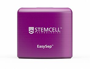
Magnet for column-free immunomagnetic separation
Cell separation buffer
EasySep™人γ / δ T细胞分离试剂盒使用EasySep™人γ / δ T细胞分离试剂盒,通过免疫磁阴性选择,从新鲜或先前冷冻的人外周血单个核细胞(PBMCs)或裂解的白细胞分离样品中轻松高效地分离高度纯化的人γ / δ T细胞。EasySep™在已发表的研究中广泛使用了20多年,它结合了单克隆抗体的特异性和无柱磁系统的简单性。
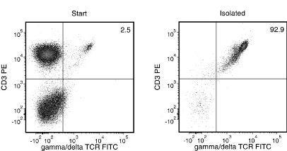
在这个EasySep™阴性选择程序中,用抗体复合物和磁性颗粒标记不需要的细胞(包括CD16和CD25细胞)。然后使用EasySep™磁铁将磁性标记的细胞与未接触的所需γ / δ T细胞分离,并将所需细胞倒入或移液到新管中。磁性细胞分离后,所需的γ / δ T细胞可用于流式细胞术、培养或DNA/RNA提取等下游应用。
了解更多关于免疫磁性的知识EasySep™技术工作原理或如何完全自动化免疫磁细胞分离RoboSep™. 或者,选择现成的、道德来源的、主要的人外周血γ δ T细胞,冷冻使用EasySep™人γ / δ T细胞分离试剂盒进行分离。探索额外的产品针对您的工作流程进行了优化,包括培养基、补充剂、抗体等。
Magnet Compatibility
Find supporting information and directions for use in the Product Information Sheet or explore additional protocols below.
This product is designed for use in the following research area(s) as part of the highlighted workflow stage(s). Explore these workflows to learn more about the other products we offer to support each research area.
| Species | Human |
|---|---|
| Magnet Compatibility | • EasySep™ Magnet (Catalog #18000) • “The Big Easy” EasySep™ Magnet (Catalog #18001) • Easy 50 EasySep™ Magnet (Catalog #18002) • RoboSep™-S (Catalog #21000) |
| Sample Source | Leukapheresis, PBMC |
| Selection Method | Negative |
细胞解离试剂
冻存的人原代细胞
小鼠(BALB/c)抗人、黑猩猩CD3单克隆IgG1抗体