产品号 #100-0139_C
小鼠单克隆抗体,抗人,恒河猴和食蟹猴CD63,未偶联
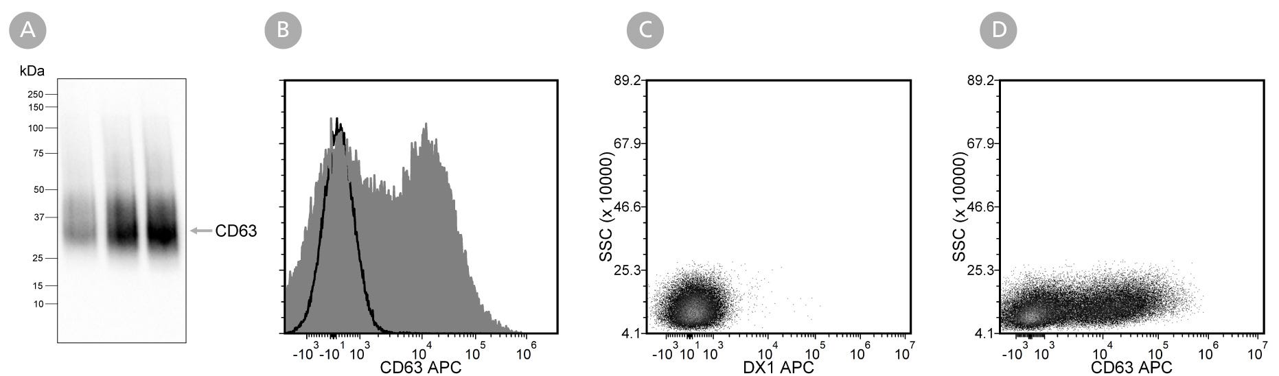
克隆H5C6抗体可识别人类CD63分子的胞外表位。CD63是一种30-60 kDa的III型溶酶体糖蛋白,属于四跨膜蛋白超家族成员。该蛋白表达广泛,存在于单核细胞、巨噬细胞、激活的嗜碱性粒细胞、成纤维细胞、平滑肌细胞及激活的血小板表面。CD63主要定位于晚期内体、溶酶体和分泌囊泡中,并在这些区室间循环转运,同时也是细胞外囊泡的特征性标志物。
四跨膜蛋白(如CD63)具有四个跨膜结构域、两个胞外环及较短的胞内N端和C端。CD63能与多种整合素、共受体及其他蛋白结合,在质膜上形成称为"四跨膜蛋白富集微域"的多分子复合物。该蛋白参与细胞活化、粘附、分化及肿瘤侵袭等多种生理过程。研究表明,CD63与肿瘤进展密切相关,其蛋白缺陷可导致Hermansky-Pudlak综合征(一种以血小板功能障碍和溶酶体贮积缺陷为特征的罕见常染色体隐性遗传病)。
亚型
一抗
靶抗原
CD63
别名
gp55
活性物种
African Green Monkey,Baboon,Capuchin Monkey,Chimpanzee,Cynomolgus,人,Rhesus
偶联
未偶联的
宿主物种
小鼠
细胞类型
其它细胞系
应用
流式细胞术,Western印迹
研究领域
细胞外囊泡研究
克隆
H5C6
基因编号
967
同种型
IgG1,kappa
Find supporting information and directions for use in the Product Information Sheet or explore additional protocols below.
This product is designed for use in the following research area(s) as part of the highlighted workflow stage(s). Explore these workflows to learn more about the other products we offer to support each research area.
| Clone | H5C6 |
|---|---|
| Gene Id | 967 |
| Alternative Names | gp55 |
| Isotype | IgG1, kappa |

免疫磁珠正选实现人细胞外囊泡快速简便分离

利用免疫磁珠正选快速简便分离人细胞外囊泡