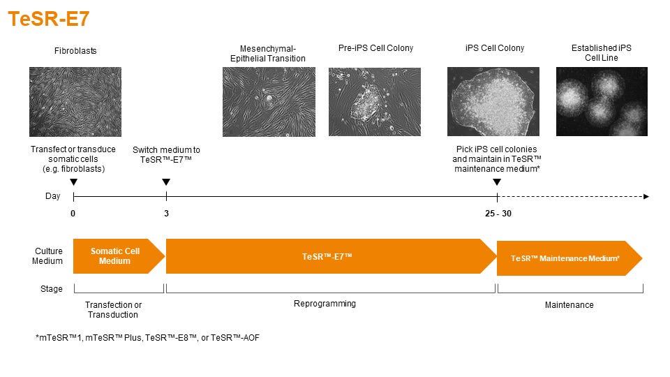
TeSR™-E7™ can be used during the entire induction phase of reprogramming (day 3 to 25+). Following reprogramming, iPS cell colonies can be isolated and propogated in feeder-free maintenance systems (eg. mTeSR™1 or TeSR™-E8™ media on Corning® Matrigel® or Vitronectin XF™ matrices).
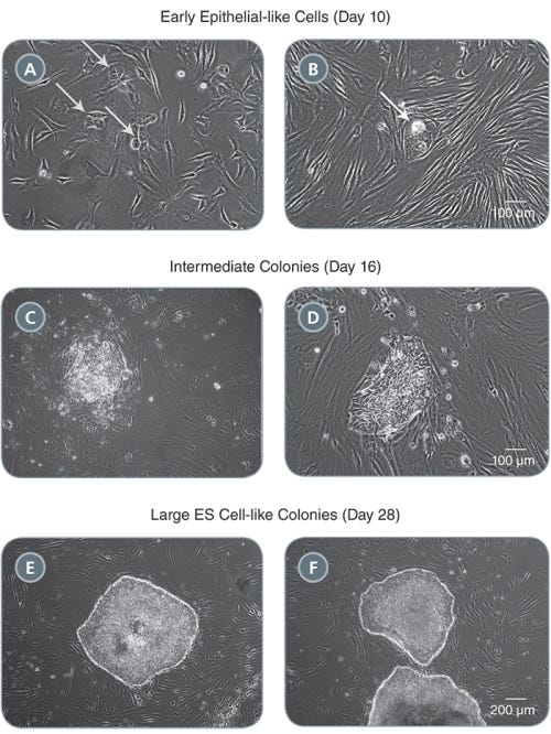
Figure 2. Morphology of Representative iPS Cell Colonies Arising During the Induction Period in TeSR™-E7™
(A-B) Small clusters of colonies with an epithelial-like morphology will appear by one to two weeks following induction (see arrows). (C-D) These clusters expand into pre-iPS cell colonies by two to three weeks. (E-F) Larger ES cell-like colonies are clearly identifiable by three to four weeks. Representative colonies from adult human fibroblasts reprogrammed with episomal vectors containing OCT-4, SOX2, KLF-4, and L-MYC are shown.
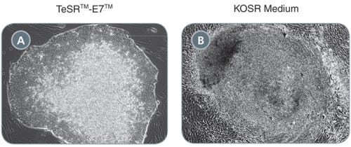
Figure 3. Comparison of Primary iPS Cell Colonies Derived Using TeSR™-E7™ and KOSR-Based Medium
(A) TeSR™-E7™ generates colonies with defined borders and less overgrowth of background fibroblasts compared to (B) KOSR-based iPS cell induction medium. Representative colonies from adult human fibroblasts reprogrammed with episomal vectors containing OCT-4, SOX2, KLF-4, and L-MYC are shown.
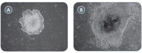
Figure 4. Comparison of Primary iPS Cell Colonies Derived Using TeSR™-E7™ with Qualified vs Unqualified bFGF
(A) TeSR™-E7™ yields easily recognizable iPS cell colonies with defined borders. (B) Unqualified components can result in colonies that have poorly defined edges and higher levels of differentiation. Representative colonies from adult human fibroblasts reprogrammed with episomal vectors containing OCT-4, SOX2, KLF-4, and L-MYC are shown.
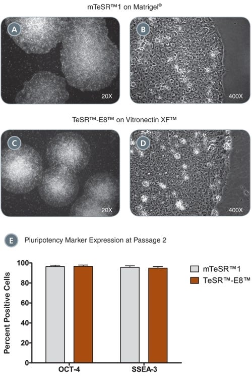
Figure 5. iPS Colonies Expanded in mTeSR™ or TeSR™-E8™
(A - D) iPS cell colonies generated in TeSR™-E7™ and expanded in either mTeSR™1 on Corning® Matrigel® (A-B) or TeSR™-E8™ on Vitronectin XF™ (C, D) exhibit classic ES cell morphology with dense colony centers, defined borders, prominent nucleoli and high nuclear-to-cytoplasmic ratios. (E) iPS cells express high levels of pluripotency markers after just two passages in either mTeSR™1 or TeSR™-E8™ as demonstrated by OCT-4 and SSEA-3 flow cytometry analysis. Data are expressed as mean ± SEM, n = 4.
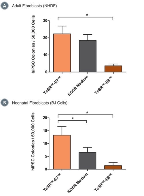
Figure 6. TeSR™-E7™ Supports Reprogramming of Human Cell Types Including Adult Dermal Fibroblasts and Neonatal Fibroblasts
Reprogramming of (A) adult normal human dermal fibroblasts (NHDF, 33 year-old female) and (B) neonatal foreskin fibroblasts (BJ cells) with episomal reprogramming vectors are shown. TeSR™-E7™ demonstrated similar (in NHDF) or greater (in BJ cells) reprogramming efficiencies compared to KOSR-based iPS cell induction medium. TeSR™-E7™ demonstrated higher reprogramming efficiencies compared to TeSR™-E8™. Data are expressed as mean ± SEM, n ≥ 6, * p ≤ 0.05.
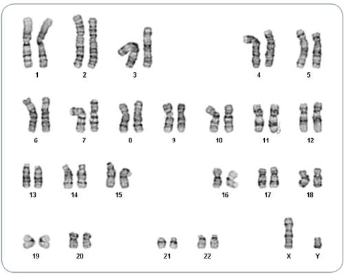
Figure 7. iPS Cells Derived in TeSR™-E7™ Display Normal Karyotype
iPS cell lines were generated in TeSR™-E7™ medium, maintained in mTeSR™1 or TeSR™-E8™ media for a minimum of 5 passages and karyotyped by G-banding karyotype analysis. Three iPS cell lines were analyzed and all demonstrated a normal karyotype; a representative karyogram is shown.
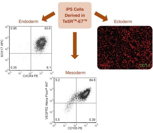
Figure 8. Directed Differentiation of iPS Cells to All Three Germ Layers
TeSR™-E7™-derived iPS cells were differentiated into all three germ layers. Endoderm specification was achieved using the STEMdiff™ Definitive Endoderm Kit, results demonstrated 93.6% SOX17 + CXCR4 + cells. Mesoderm specification was demonstrated using a STEMdiff™ APEL™ medium-based endothelial differentiation protocol, results demonstrated &ht;99% CD31 + cells (data not shown) and 84.8% VEGFR2 + CD105 + cells. Ectoderm specification was demonstrated using STEMdiff™ Neural Induction Medium, immunocytochemistry shows high levels of PAX6 staining with no detectable OCT-4 staining by day 9 of neural induction.
This product is designed for use in the following research area(s) as part of the highlighted workflow stage(s). Explore these workflows to learn more about the other products we offer to support each research area.
Disruption of GRIN2B Impairs Differentiation in Human Neurons. S. Bell et al. Stem cell reports 2018 JUL
Abstract
Heterozygous loss-of-function mutations in GRIN2B, a subunit of the NMDA receptor, cause intellectual disability and language impairment. We developed clonal models of GRIN2B deletion and loss-of-function mutations in a region coding for the glutamate binding domain in human cells and generated neurons from a patient harboring a missense mutation in the same domain. Transcriptome analysis revealed extensive increases in genes associated with cell proliferation and decreases in genes associated with neuron differentiation, a result supported by extensive protein analyses. Using electrophysiology and calcium imaging, we demonstrate that NMDA receptors are present on neural progenitor cells and that human mutations in GRIN2B can impair calcium influx and membrane depolarization even in a presumed undifferentiated cell state, highlighting an important role for non-synaptic NMDA receptors. It may be this function, in part, which underlies the neurological disease observed in patients with GRIN2B mutations.
Thank you for your interest in IntestiCult™ Organoid Growth Medium (Human). Please provide us with your contact information and your local representative will contact you with a customized quote. Where appropriate, they
can also assist you with a(n):
Estimated delivery time for your area
Product sample or exclusive offer
In-lab demonstration
By submitting this form, you are providing your consent to STEMCELL Technologies Canada Inc. and its subsidiaries and affiliates (“STEMCELL”) to collect and use your information, and send you newsletters and emails in accordance with our
privacy policy. Please contact us with any questions that you may have. You can unsubscribe or change your email preferences at any time.
Legal Statement: This product was developed under license to intellectual property owned by WiCell™ Research Institute. This product is sold for research use only (whether the buyer is an academic or for-profit entity) under a non-transferable, limited-use license. Purchase of this product does not include the right to sell, use or otherwise transfer this product for commercial purposes (i.e., any activity undertaken for consideration, such as use of this product for manufacturing, or resale of this product or any materials made using this product, or use of this product or any materials made using this product to provide services) or clinical use (i.e., administration of this product or any material using this product to humans) or the right to implant any material made using this product into an animal by, or in collaboration with, a for-profit entity, for purposes other than basic pre-clinical research applications (including without limitation teratoma assays) to validate the function of the cells. Purchasers who do not agree to the terms and conditions set forth above should return the product in acceptable conditions to the seller for a refund. PRODUCTS ARE FOR RESEARCH USE ONLY AND NOT INTENDED FOR HUMAN OR ANIMAL DIAGNOSTIC OR THERAPEUTIC USES UNLESS OTHERWISE STATED. FOR ADDITIONAL INFORMATION ON QUALITY AT STEMCELL, REFER TO WWW.STEMCELL.COM/COMPLIANCE.