翻译:PneumaCult™-Ex Plus Medium

无血清和无bpe培养基用于原代人气道上皮细胞的扩增

产品号 #(选择产品)

产品号 #05040_C

无血清和无bpe培养基用于原代人气道上皮细胞的扩增

产品优势

  • 一种明确的,无血清和无bpe的细胞培养基,提供一致的性能
  • 与其他市售的扩增介质相比,PneumaCult™-Ex Plus Medium在每次传代中支持更多的细胞扩增
  • 当与PneumaCult™-ALI Medium或PneumaCult™-ALI- s Medium一起使用时,与其他市售扩展介质相比,即使在延长传代后,PneumaCult™-Ex Plus Medium也能支持更好的ALI分化潜力

产品组分包括

  • PneumaCult™-Ex Plus基础培养基,490毫升
  • PneumaCult™-Ex Plus 50X补充剂,10毫升
Interested in trying STEMCELL's products for respiratory research? Fill out this form to request information about introductory offers.
Products for Your Protocol
To see all required products for your protocol, please consult the Protocols and Documentation.

概述

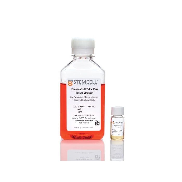
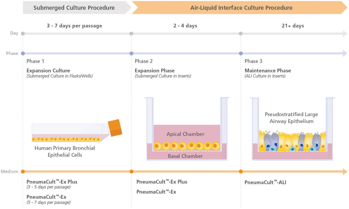
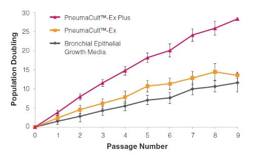
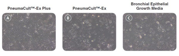
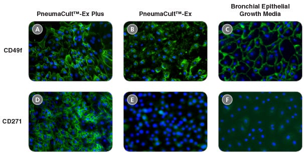
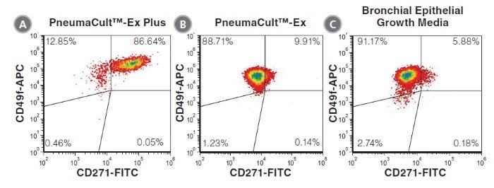
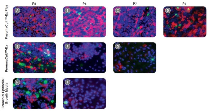
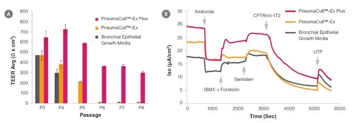
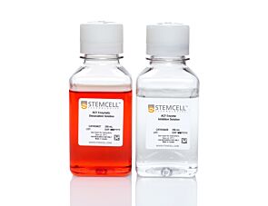
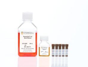
PneumaCult™- ex Plus Medium是一种明确的,不含血清和bpe的细胞培养基,与其他市售的扩增培养基相比,它在每次传代时支持更多的人气道和鼻上皮细胞扩增。该培养基还支持至少两个额外的细胞扩增通道,具有更好的分化潜力,定义为使用PneumaCult™-ALI培养基(目录#05001)在气液界面(ALI)形成假层状粘膜纤毛上皮或使用PneumaCult™-ALI- s培养基(目录#05050)形成立方上皮的能力。

PneumaCult™-Ex Plus和PneumaCult™-ALI或PneumaCult™-ALI- s构成了一个完全集成的无bpe培养系统,用于体外人体气道建模。这一强大而明确的系统是基础呼吸研究、毒性研究和药物开发的宝贵工具。

了解如何培养人类气道上皮细胞在我们的ALI按需肺课程或浏览我们的常见问题(FAQs)关于使用PneumaCult™的ALI培养工作流程。

PneumaCult™-ALI 培养基和PneumaCult™-Ex Plus培养基(产品号 #05040)共同构成了一个一体化的无 BPE 的体外人呼吸道培养模型。该培养模型效果稳定且成分明确,是呼吸系统基础研究、毒性研究和药物开发的宝贵工具。

通过我们的点播式肺部课程学习如何在气液界面培养人呼吸道上皮细胞,或浏览关于使用PneumaCult™进行气液界面培养的常见问题解答(FAQ)。

Subtype
Specialized Media
 
Cell Type
Airway Cells
 
Species
Human
 
Application
Cell Culture, Expansion, Maintenance
 
Brand
PneumaCult
 
Area of Interest
Epithelial Cell Biology
 
Formulation Category
Serum-Free
 

Data Figures

Figure 1. Overview of the PneumaCult™ culture system

Expansion of human bronchial epithelial cells (HBECs) in submerged culture is performed with PneumaCult™-Ex Plus or PneumaCult™-Ex. During the early “Expansion Phase” of the air-liquid interface (ALI) culture procedure, PneumaCult™-Ex Plus or PneumaCult™-Ex is applied to the apical and basal chambers. Upon reaching confluence, the culture is air-lifted by removing the culture medium from both chambers, and adding PneumaCult™-ALI to the basal chamber only. Differentiation into a pseudostratified mucociliary epithelium is obtained following 21-28 days of incubation and can be maintained for more than one year.

Figure 2. HBECs cultured in PneumaCult™-Ex Plus have a faster expansion rate compared to those cultured in PneumaCult™-Ex and Bronchial Epithelial Growth Media

Commercially available, cryopreserved P1 HBECs were seeded into PneumaCult™-Ex Plus, PneumaCult™-Ex, or Bronchial Epithelial Growth Media. Cells cultured in PneumaCult™-Ex Plus have a significantly higher proliferation rate over 9 passages compared to those maintained in either control medium (n=6).

Figure 3. Representative morphology of HBECs

Representative live culture images for P4 HBECs cultured in PneumaCult™-Ex Plus, PneumaCult™-Ex, or Bronchial Epithelial Growth Media. Cells cultured in PneumaCult™-Ex Plus (A) are smaller and more tightly packed than those cultured in PneumaCult™-Ex (B) or Bronchial Epithelial Growth Media (C). All images were taken using a 10X objective.

Figure 4. HBECs cultured in PneumaCult™-Ex Plus maintain widespread expression of the basal cell markers CD49f and CD271

Immunocytochemistry detection of basal cell markers - CD49f (A, B, and C) and CD271 (D, E, and F) - for P4 HBECs cultured in PneumaCult™-Ex Plus (A and D), PneumaCult™-Ex (B and E), and Bronchial Epithelial Growth Media (C and F). All images were taken using a 10X objective.

Figure 5. HBECs cultured in PneumaCult™-Ex Plus have a higher proportion of CD271+CD49f+ cells

P4 HBECs cultured in PneumaCult™-Ex Plus (A), PneumaCult™-Ex (B), and Bronchial Epithelial Growth Media (C) were characterized by flow cytometry to detect expression of the basal cell markers CD49f and CD271. HBECs cultured in PneumaCult™-Ex Plus (A) have a higher proportion of cells coexpressing CD49f and CD271, compared to those cultured in PneumaCult™-Ex (B) and Bronchial Epithelial Growth Media (C).

Figure 6. HBECs cultured in PneumaCult™-Ex Plus differentiate into a pseudostratified mucociliary epithelium at later passages with the use of PneumaCult™-ALI

P4 HBECs were seeded and passaged using PneumaCult™-Ex Plus, PneumaCult™-Ex, or Bronchial Epithelial Growth Media, followed by ALI differentiation at each passage (P5-8) with the use of PneumaCult™-ALI. The ALI cultures at 28 days post air-lift were fixed and stained with antibodies for cilia marker AC-tubulin (red) and the goblet cell marker Muc5AC (green). The nuclei are counterstained with DAPI (blue). All images were taken using a 20X objective.

Figure 7. Electrophysiological characterization of differentiated HBECs (P4) that were expanded in PneumaCult™-Ex Plus, PneumaCult™-Ex, and Bronchial Epithelial Growth Media

Transepithelial electrical resistance (TEER) (A) and representative characterization of the ion channel activities (B) for ALI cultures at 28 days post air-lift using HBECs expanded in PneumaCult™-Ex Plus, PneumaCult™-Ex, or Bronchial Epithelial Growth Media. Amiloride: ENaC inhibitor. IBMX and Forskolin: CFTR activators. Genistein: CFTR potentiator. CFTRinh-172: CFTR inhibitor. UTP: Calciumactivated Chloride channels (CaCCs) activator. All ALI differentiation cultures were performed using PneumaCult™-ALI.

Protocols and Documentation

Find supporting information and directions for use in the Product Information Sheet or explore additional protocols below.

Document Type
Product Name
Catalog #
Lot #
Language
Catalog #
05040
Lot #
All
Language
English
Document Type
Safety Data Sheet 1
Catalog #
05040
Lot #
All
Language
English
Document Type
Safety Data Sheet 2
Catalog #
05040
Lot #
All
Language
English

Applications

This product is designed for use in the following research area(s) as part of the highlighted workflow stage(s). Explore these workflows to learn more about the other products we offer to support each research area.

Resources and Publications

Educational Materials (17)

Publications (14)

M1-like, but not M0- or M2-like, macrophages, reduce RSV infection of primary bronchial epithelial cells in a media-dependent fashion. N. J. Ronaghan et al. PloS one 2022

Abstract

Respiratory syncytial virus (RSV) is a common childhood infection that in young infants can progress into severe bronchiolitis and pneumonia. Disease pathogenesis results from both viral mediated and host immune processes of which alveolar macrophages play an important part. Here, we investigated the role of different types of alveolar macrophages on RSV infection using an in vitro co-culture model involving primary tissue-derived human bronchial epithelial cells (HBECs) and human blood monocyte-derived M0-like, M1-like, or M2-like macrophages. It was hypothesized that the in vitro model would recapitulate previous in vivo findings of a protective effect of macrophages against RSV infection. It was found that macrophages maintained their phenotype for the 72-hour co-culture time period and the bronchial epithelial cells were unaffected by the macrophage media. HBEC infection with RSV was decreased by M1-like macrophages but enhanced by M0- or M2-like macrophages. The medium used during the co-culture also impacted the outcome of the infection. This work demonstrates that alveolar macrophage phenotypes may have differential roles during epithelial RSV infection, and demonstrates that an in vitro co-culture model could be used to further investigate the roles of macrophages during bronchial viral infection.
Effect of apical chloride concentration on the measurement of responses to CFTR modulation in airway epithelia cultured from nasal brushings. P. E. Bratcher et al. Physiological reports 2020 oct

Abstract

INTRODUCTION One method for assessing the in vitro response to CFTR-modulating compounds is by analysis of epithelial monolayers in an Ussing chamber, where the apical and basolateral surfaces are isolated and the potential difference, short-circuit current, and transepithelial resistance can be monitored. The effect of a chloride ion gradient across airway epithelia on transepithelial chloride transport and the magnitude of CFTR modulator efficacy were examined. METHODS CFTR-mediated changes in the potential difference and transepithelial currents of primary human nasal epithelial cell cultures were quantified in Ussing chambers with either symmetrical solutions or reduced chloride solutions in the apical chamber. CFTR activity in homozygous F508del CFTR epithelia was rescued by treatment with VX-661, C4/C18, 4-phenylbutyrate (4-PBA) for 24 hr at 37°C or by incubation at 29°C for 48 hr. RESULTS Imposing a chloride gradient increased CFTR-mediated and CaCC-mediated ion transport. Treatment of F508del CFTR homozygous cells with CFTR modulating compounds increased CFTR activity, which was significantly more evident in the presence of a chloride gradient. This observation was recapitulated with temperature-mediated F508del CFTR correction. CONCLUSIONS Imposing a chloride gradient during Ussing chamber measurements resulted in increased CFTR-mediated ion transport in expanded non-CF and F508del CFTR homozygous epithelia. In F508del CFTR homozygous epithelia, the magnitude of response to CFTR modulating compounds or low temperature was greater when assayed with a chloride gradient compared to symmetrical chloride, resulting in an apparent increase in measured efficacy. Future work may direct which methodologies utilized to quantify CFTR modulator response in vitro are most appropriate for the estimation of in vivo efficacy.
A Revised Protocol for Culture of Airway Epithelial Cells as a Diagnostic Tool for Primary Ciliary Dyskinesia. J. L. Coles et al. Journal of clinical medicine 2020 nov

Abstract

Air-liquid interface (ALI) culture of nasal epithelial cells is a valuable tool in the diagnosis and research of primary ciliary dyskinesia (PCD). Ex vivo samples often display secondary dyskinesia from cell damage during sampling, infection or inflammation confounding PCD diagnostic results. ALI culture enables regeneration of healthy cilia facilitating differentiation of primary from secondary ciliary dyskinesia. We describe a revised ALI culture method adopted from April 2018 across three collaborating PCD diagnostic sites, including current University Hospital Southampton COVID-19 risk mitigation measures, and present results. Two hundred and forty nasal epithelial cell samples were seeded for ALI culture and 199 (82.9{\%}) were ciliated. Fifty-four of 83 (63.9{\%}) ex vivo samples which were originally equivocal or insufficient provided diagnostic information following in vitro culture. Surplus basal epithelial cells from 181 nasal brushing samples were frozen in liquid nitrogen; 39 samples were ALI-cultured after cryostorage and all ciliated. The ciliary beat patterns of ex vivo samples (by high-speed video microscopy) were recapitulated, scanning electron microscopy demonstrated excellent ciliation, and cilia could be immuno-fluorescently labelled (anti-alpha-tubulin and anti-RSPH4a) in representative cases that were ALI-cultured after cryostorage. In summary, our ALI culture protocol provides high ciliation rates across three centres, minimising patient recall for repeat brushing biopsies and improving diagnostic certainty. Cryostorage of surplus diagnostic samples was successful, facilitating PCD research.

更多信息

更多信息
Species Human
Formulation Category Serum-Free
Legal Statement: This product was developed under a license to intellectual property owned or controlled by Propagenix, Inc. This product is sold for research use only (which includes pre-clinical research) under a non-transferable, limited-use license. Purchase of this product does not include the right to sell, use or otherwise transfer this product for commercial purposes or clinical use. Purchasers wishing to use the product for purposes other than research use should contact Propagenix, Inc. (www.propagenix.com/about#contact-us). PRODUCTS ARE FOR RESEARCH USE ONLY AND NOT INTENDED FOR HUMAN OR ANIMAL DIAGNOSTIC OR THERAPEUTIC USES UNLESS OTHERWISE STATED. FOR ADDITIONAL INFORMATION ON QUALITY AT STEMCELL, REFER TO WWW.STEMCELL.COM/COMPLIANCE.
版权 © 2025 STEMCELL Technologies 技术有限公司。保留所有权利。