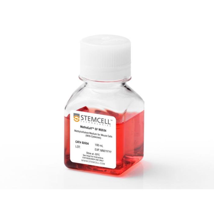
产品号 #03534_C
含重组细胞因子(不含促红细胞生成素[EPO])的甲基纤维素培养基用于小鼠骨髓祖细胞
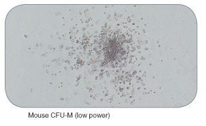
MethoCult™GF M3534针对小鼠骨髓、脾脏、外周血和胎儿肝细胞的集落形成单位(CFU)检测中粒细胞-巨噬细胞祖细胞(CFU- gm、CFU- g、CFU- m)的生长和计数进行了优化。MethoCult™M3534不支持红细胞祖细胞(BFU-E和CFU-E)的生长,因为它不含促红细胞生成素(EPO)。该制剂与STEMvision™软件兼容,用于小鼠骨髓CFU检测的自动菌落计数。
浏览我们的常见问题(FAQs)进行CFU化验。
Contains
• Methylcellulose in Iscove's MDM
• Fetal bovine serum
• Bovine serum albumin
• Recombinant human insulin
• Human transferrin (iron-saturated)
• 2-Mercaptoethanol
• Recombinant mouse stem cell factor (SCF)
• Recombinant mouse interleukin 3 (IL-3)
• Recombinant human interleukin 6 (IL-6)
• Supplements
Subtype
Semi-Solid Media, Specialized Media
Cell Type
Hematopoietic Stem and Progenitor Cells
Species
Mouse
Application
Cell Culture, Colony Assay, Functional Assay
Brand
MethoCult
Area of Interest
Drug Discovery and Toxicity Testing, Stem Cell Biology
Formulation Category
Methylcellulose-Based
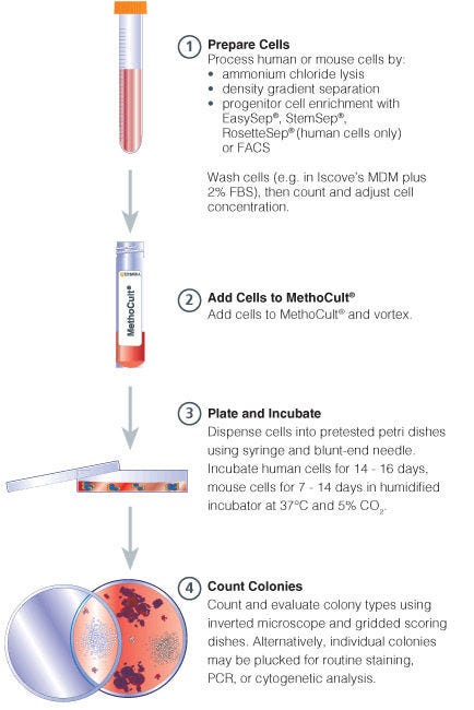
Find supporting information and directions for use in the Product Information Sheet or explore additional protocols below.
This product is designed for use in the following research area(s) as part of the highlighted workflow stage(s). Explore these workflows to learn more about the other products we offer to support each research area.
| Species | Mouse |
|---|---|
| Contains | • Methylcellulose in Iscove's MDM • Fetal bovine serum • Bovine serum albumin • Recombinant human insulin • Human transferrin (iron-saturated) • 2-Mercaptoethanol • Recombinant mouse stem cell factor (SCF) • Recombinant mouse interleukin 3 (IL-3) • Recomb |
| Formulation Category | Methylcellulose-Based |

用于造血集落形成单位(CFU)测定的自动化和标准化集落计数
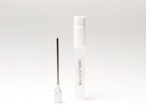
16号鲁尔锁针头
用于精确分装甲基纤维素培养基