MesenCult™ 成骨细胞分化试剂盒 (人)

用于体外诱导人MSC分化为成骨细胞

产品号 #(选择产品)

产品号 #05465_C

用于体外诱导人MSC分化为成骨细胞

产品优势

  • 兼容先前在MesenCult™扩增培养基中培养扩增的人MSC。
  • 即用型双组分试剂盒
  • 严格的原材料筛选和质量控制可确保最低的批次间的差异性。

产品组分包括

  • MesenCult™成骨分化基础培养基(人),200 mL
  • MesenCult™成骨分化5X补充剂(人),50ml

概述

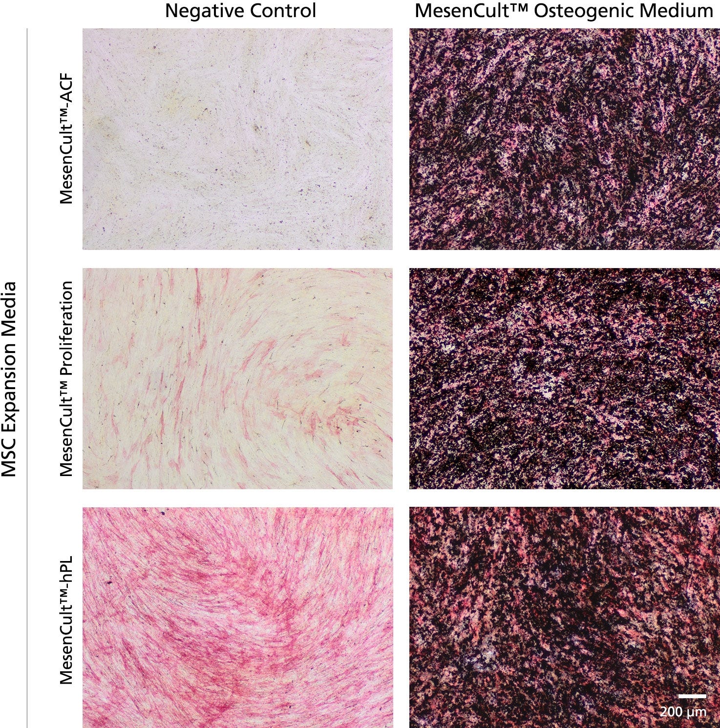
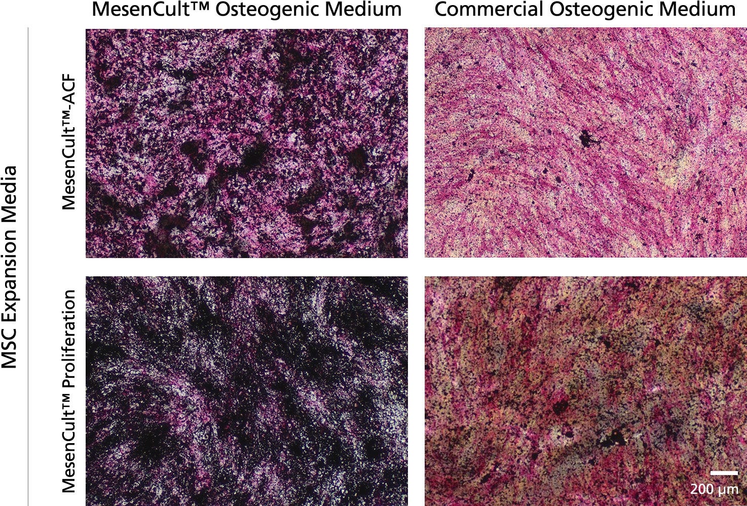
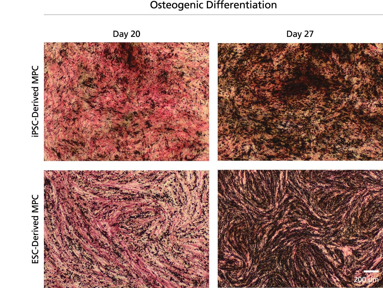
MesenCult™成骨分化试剂盒(人)是专门为原代人间充质基质细胞和hpsc衍生的间充质祖细胞(也称为间充质干细胞或MSCs)在体外分化成成骨谱系的细胞而制定的。该试剂盒适用于人骨髓(BM)或脂肪来源的间充质干细胞的分化,之前在含血清培养基中培养扩增(例如MesenCult™增殖试剂盒[人;目录#05411]或MesenCult™-hPL Medium[人类;目录#05439])或无动物成分的MesenCult™-ACF Plus Medium[目录#05445])。

Cell Type
Mesenchymal Stem and Progenitor Cells, Osteoblasts
 
Application
Cell Culture, Differentiation
 
Brand
MesenCult
 
Area of Interest
Stem Cell Biology
 

Data Figures

Figure 1. Robust Bone Marrow Mesenchymal Stem and Progenitor Cells (BM MSCs) Osteogenic Differentiation is Achieved in 14 days

Human BM MSCs were derived and expanded for 3 passages using the MesenCult™-ACF Culture Kit (Catalog #05449), MesenCult™ Proliferation Kit (Catalog #05411) or in MesenCult™-hPL Medium (05439). Once MSCs reach greater than 95% confluency, MesenCult™ Osteogenic medium was added to each MSC culture. Osteogenic differentiation was observed within 14 days of induction as indicated by strong alkaline phosphatase activity (red stain) and bone mineralization by the von Kossa method (black stain). Negative controls of undifferentiated MSC cultures were kept in each MesenCult™ MSC expansion media for the same time period. Negative Controls show little or no alkaline phosphatase activity and bone mineralization.

Figure 2. MesenCult™ Osteogenic Medium Leads to Faster and Stronger Osteogenic Differentiation When Compared to A Commercial Osteogenic Differentiation Medium

BM MSCs derived and expanded in MesenCult™-ACF or MesenCult™ Proliferation medium were differentiated for 14 days in either MesenCult™ Osteogenic or another Commercial Osteogenic Medium. Differentiation assays using the MesenCult™ Osteogenic medium displayed stronger alkaline phosphatase activity (red stain) and bone mineralization (black stain) when compared to cultures differentiated with a Commercial Osteogenic Medium.

Figure 3. Osteogenic Differentiation of ES-Derived Mesenchymal Progenitor Cells (MPCs)

Mesenchymal progenitor cells (MPCs) were derived from a human iPS or ES cell line using the STEMdiff™ Mesenchymal Progenitor Kit (Catalog #05240) and expanded for 18 or 17 passages, respectively. Cultures of iPS- and ES-derived MPCs were then differentiated for 20 or 27 days in MesenCult™ Osteogenic Differentiation Medium. Strong alkaline phosphatase activity (red stain) and bone mineralization (black stain) were observed at 20 days of osteogenic differentiation, which was further enhanced after 27 days of osteogenic differentiation.

Protocols and Documentation

Find supporting information and directions for use in the Product Information Sheet or explore additional protocols below.

Document Type
Product Name
Catalog #
Lot #
Language
Catalog #
05465
Lot #
All
Language
English
Document Type
Safety Data Sheet 1
Catalog #
05465
Lot #
All
Language
English
Document Type
Safety Data Sheet 2
Catalog #
05465
Lot #
All
Language
English

Applications

This product is designed for use in the following research area(s) as part of the highlighted workflow stage(s). Explore these workflows to learn more about the other products we offer to support each research area.

Resources and Publications

Educational Materials (6)

Publications (9)

In situ repair abilities of human umbilical cord-derived mesenchymal stem cells and autocrosslinked hyaluronic acid gel complex in rhesus monkeys with intrauterine adhesion. L. Wang et al. Science advances 2020 may

Abstract

Increasing occurrence of moderate to severe intrauterine adhesion (IUA) is seriously affecting the quality of human life. The aim of the study was to establish IUA models in nonhuman primates and to explore the dual repair effects of human umbilical cord-derived mesenchymal stem cells (huMSCs) loaded on autocrosslinked hyaluronic acid gel (HA-GEL) on endometrial damage and adhesion. Here, we recorded the menstrual cycle data in detail with uterine cavities observed and endometrial tissues detected after intervention, and the thicker endometria, decreased amount of fibrotic formation, increased number of endometrium glands, etc., suggested that both HA-GEL and huMSC/HA-GEL complexes could partially repair IUA caused by mechanical injury, but huMSC/HA-GEL complex transplantation had notable dual repair effects: a reliable antiadhesion property and the promotion of endometrial regeneration.
Altered Surface Hydrophilicity on Copolymer Scaffolds Stimulate the Osteogenic Differentiation of Human Mesenchymal Stem Cells. Z. Xing et al. Polymers 2020 jun

Abstract

BACKGROUND Recent studies have suggested that both poly(l-lactide-co-1,5-dioxepan-2-one) (or poly(LLA-co-DXO)) and poly(l-lactide-co-$\epsilon$-caprolactone) (or poly(LLA-co-CL)) porous scaffolds are good candidates for use as biodegradable scaffold materials in the field of tissue engineering; meanwhile, their surface properties, such as hydrophilicity, need to be further improved. METHODS We applied several different concentrations of the surfactant Tween 80 to tune the hydrophilicity of both materials. Moreover, the modification was applied not only in the form of solid scaffold as a film but also a porous scaffold. To investigate the potential application for tissue engineering, human bone marrow mesenchymal stem cells (hMSCs) were chosen to test the effect of hydrophilicity on cell attachment, proliferation, and differentiation. First, the cellular cytotoxicity of the extracted medium from modified scaffolds was investigated on HaCaT cells. Then, hMSCs were seeded on the scaffolds or films to evaluate cell attachment, proliferation, and osteogenic differentiation. The results indicated a significant increasing of wettability with the addition of Tween 80, and the hMSCs showed delayed attachment and spreading. PCR results indicated that the differentiation of hMSCs was stimulated, and several osteogenesis related genes were up-regulated in the 3{\%} Tween 80 group. Poly(LLA-co-CL) with 3{\%} Tween 80 showed an increased messenger Ribonucleic acid (mRNA) level of late-stage markers such as osteocalcin (OC) and key transcription factor as runt related gene 2 (Runx2). CONCLUSION A high hydrophilic scaffold may speed up the osteogenic differentiation for bone tissue engineering.
Human Umbilical Cord Mesenchymal Stem Cells Attenuate Abdominal Aortic Aneurysm Progression in Sprague-Dawley Rats: Implication of Vascular Smooth Muscle Cell Phenotypic Modulation. H. Wen et al. Stem cells and development 2020 jul

Abstract

Abdominal aortic aneurysm (AAA) is life-threatening, for which efficient nonsurgical treatment strategy has not been available so far. Several previous studies investigating the therapeutic effect of mesenchymal stem cells (MSCs) in AAA indicated that MSCs could inhibit aneurysmal inflammatory responses and extracellular matrix destruction, and suppress aneurysm occurrence and expansion. Vascular smooth muscle cell (VSMC) phenotypic plasticity is reported to be predisposed in AAA initiation and progression. However, little is known about the effect of MSCs on VSMC phenotypic modulation in AAA. In this study, we investigate the therapeutic efficacy of umbilical cord mesenchymal stem cells (UC-MSCs) in elastase-induced AAA model and evaluate the effect of UC-MSC on VSMC phenotypic regulation. We demonstrate that the intravenous injection of UC-MSC attenuates elastase-induced aneurysmal expansion, reduces elastin degradation and fragmentation, inhibits MMPs and TNF-$\alpha$ expression, and preserves and/or restores VSMC contractile phenotype in AAA. Taken together, these results highlight the therapeutic and VSMC phenotypic modulation effects of UC-MSC in AAA progression, which further indicates the potential of applying UC-MSC as an alternative treatment candidate for AAA.
PRODUCTS ARE FOR RESEARCH USE ONLY AND NOT INTENDED FOR HUMAN OR ANIMAL DIAGNOSTIC OR THERAPEUTIC USES UNLESS OTHERWISE STATED. FOR ADDITIONAL INFORMATION ON QUALITY AT STEMCELL, REFER TO WWW.STEMCELL.COM/COMPLIANCE.
版权 © 2025 STEMCELL Technologies 技术有限公司。保留所有权利。