EasySep™人NK细胞分选试剂盒

不带标记的人NK细胞免疫磁珠负选

产品号 #100-0960

人NK细胞大规模免疫磁珠负选

产品优势

  • 操作简单、快捷,且无需分离柱
  • 纯度高达94%
  • 分选得到的细胞不带标记

产品组分包括

  • EasySep™人NK细胞分选试剂盒(产品号 #17955)
    • EasySep™人NK细胞分选抗体混合物,1 x 1 mL
    • EasySep™ Dextran RapidSpheres™磁珠,1 x 1 mL
  • EasySep™人NK细胞分选试剂盒(产品号 #100-0960)
    • EasySep™人NK细胞分选抗体混合物,1 x 10 mL(产品号 #300-0475)
    • EasySep™ Dextran RapidSpheres™磁珠,1 x 1 mL(产品号 #300-0380)
  • EasySep™人NK细胞分选试剂盒(产品号 #17955RF)
    • EasySep™人NK细胞分选抗体混合物,1 mL
    • EasySep™ Dextran RapidSpheres™磁珠,1 mL
    • EasySep™人NK细胞分选抗体混合物,1 mL
    • EasySep™ Dextran RapidSpheres™磁珠,1 mL
  • RoboSep™ 缓冲液(产品号 #20104)
      • RoboSep™过滤吸头(产品号 #20125)

What Our Scientist Says

We know that cell isolation is only part of your workflow. We designed this kit to isolate NK cells in as little as 8 minutes, so you can get to your downstream experiments as soon as possible.

Manreet ChehalScientist
Manreet Chehal
Scientist, Scientist

概述

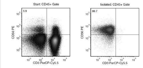
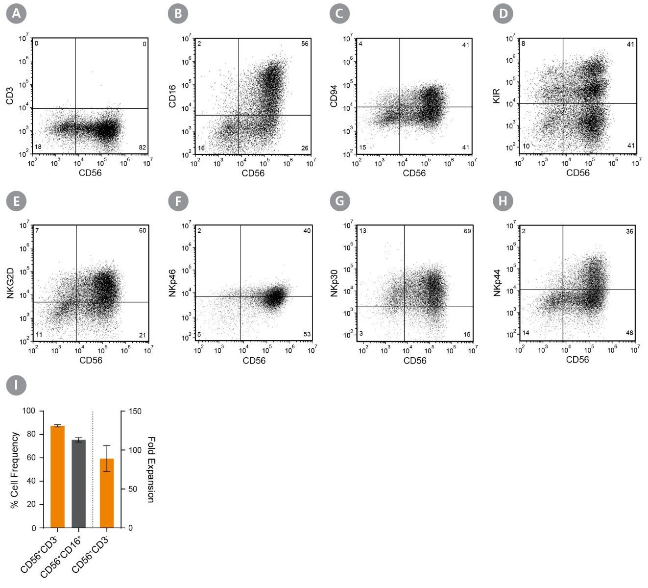
通过免疫磁珠负选,从新鲜或冻存的人外周血单个核细胞 (PBMCs) 或洗涤的白细胞单采术样本中分离出无磁珠标记和高纯度的自然杀伤(NK)细胞。EasySep™技术结合单克隆抗体的特异性和无柱磁分选系统的简便性,已在发表的研究中广泛应用超过20年。
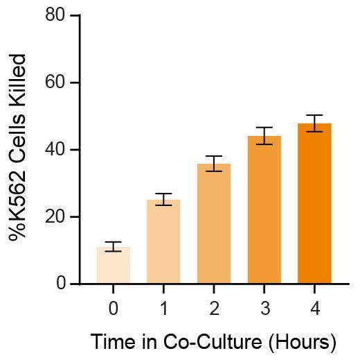
在该EasySep™负选流程中,非目的细胞通过抗体复合物与磁珠被标记。以下非目标细胞将被去除:粒细胞、T细胞、B细胞、单核细胞、树突状细胞、红细胞。通过EasySep™磁极将被磁珠标记的细胞与未被标记的目的细胞分离,接着简单地将目的细胞倾倒或吸至一个新的试管中。磁珠分选最快仅需8分钟,所得NK细胞可立即用于流式细胞术、培养或DNA/RNA提取等下游应用。

该产品可替代EasySep™人NK细胞富集试剂盒 (产品号 #19055) 进行更快地细胞分选。
针对包含CD36+和HLA-DR+新生亚群的NK细胞分选,我们推荐EasySep™人 Pan NK细胞分选试剂盒(产品号100-1580),最快8分钟即可完成分选。
如需从白细胞单采术样本中大规模分选人NK细胞,请参阅大规格(1x10^10 个细胞)的试剂盒(产品号 #100-0960)。深入了解免疫磁珠EasySep™技术原理,或探索如何通过RoboSep™实现全自动免疫磁珠细胞分选。您亦可选择即用型、符合伦理来源的原代人外周血NK细胞,该细胞通过EasySep™人NK细胞分选试剂盒新鲜分离。探索更多为您实验流程优化的其它产品,包括培养基、补充剂、抗体等。

MAGNET COMPATIBILITY
• EasySep™ Magnet (Catalog #18000)
• “The Big Easy” EasySep™ Magnet (Catalog #18001)
• Easy 50 EasySep™ Magnet (Catalog #18002)
• EasyEights™ EasySep™ Magnet (Catalog #18103)
• RoboSep™-S (Catalog #21000)
• Easy 250 EasySep™ Magnet (Catalog #100-0821)
 
SUBTYPE
Cell Isolation Kits
 
CELL TYPE
NK Cells
 
SPECIES
Human
 
SAMPLE SOURCE
Leukapheresis, PBMC
 
SELECTION METHOD
Negative
 
APPLICATION
Cell Isolation
 
BRAND
EasySep, RoboSep
 
AREA OF INTEREST
Chimerism, Immunology

实验数据

Separation of natural killer cells using EasySep™ Human NK Cell Isolation Kit

Figure 1. Typical EasySep™ Human NK Cell Isolation Profile

Starting with human PBMCs, the NK cell (CD3-CD56+) content of the isolated fraction is typically 85.0 ± 8.0% (mean ± SD). In the above example, the final purities of the start and isolated fractions are 5.9% and 86.7%, respectively.

Cell Frequency, Fold Expansion, and Phenotyping of Natural Killer (NK) Cells Cultured Using the ImmunoCult™ NK Cell Expansion Kit

Figure 2. CD56+CD3− NK Cells Expand Over 14 Days in Feeder- and Serum-Free Culture Conditions

Human CD56+CD3− NK cells isolated using EasySep™ Human NK Cell Isolation Kit (Catalog #17955) were cultured using ImmunoCult™ NK Cell Expansion Kit (Catalog #100-0711) for 14 days. Cells were harvested and analyzed for expression of characteristic NK cell markers, including CD56, CD3, CD16, CD94, KIR, NKG2D, NKp46, NKp30, and NKp44 by flow cytometry. Staining for killer cell immunoglobulin-like receptor (KIR) molecules was performed using two different antibody clones, HP-MA4 and 180704, which recognize distinct KIR molecules. Dead cells were excluded by light-scatter profile and DRAQ7™ staining. (A - H) Representative flow cytometry plots. (I) The average frequencies of viable CD56+CD3− and CD56+CD16+ NK cells on Day 14 were 87 ± 1% and 75 ± 2%, respectively. The average fold expansion of CD56+CD3− cells was 89 ± 17. Results shown represent mean ± SEM (n = 34).

Cytotoxicity of Expanded Natural Killer (NK) Cells Co-Cultured with K562 Cells

Figure 3. Expanded NK Cells Are Functional, Killing K562 Cells in Co-Culture

CD56+CD3− NK cells isolated using EasySep™ Human NK Cell Isolation Kit (Catalog #17955) were expanded using ImmunoCult™ NK Cell Expansion Kit (Catalog #100-0711) and co-cultured with Incucyte® Cytolight Rapid Dye-labeled K562 cells at a 1:1 ratio of NK:K562 cells at 37°C for 4 hours. Incucyte® Caspase-3/7 Dye, a caspase-inducible dye, was added to the co-culture to detect caspase-induced apoptosis of the K562 cells. Images were obtained every hour using the Incucyte® imaging system and then analyzed to determine % killing (# apoptotic K562 cells ÷ # total labeled K562 cells). After 4 hours, an average of 48 ± 2.4% K562 cells were killed (n = 9). Data represent mean ± SEM.

产品说明书及文档

Find supporting information and directions for use in the Product Information Sheet or explore additional protocols below.

Document Type
Product Name
Catalog #
Lot #
Language
Catalog #
17955RF
Lot #
All
Language
English
Catalog #
100-0960
Lot #
All
Language
English
Catalog #
17955
Lot #
All
Language
English
Document Type
Safety Data Sheet 1
Catalog #
17955RF
Lot #
All
Language
English
Document Type
Safety Data Sheet 2
Catalog #
17955RF
Lot #
All
Language
English
Document Type
Safety Data Sheet 3
Catalog #
17955RF
Lot #
All
Language
English
Document Type
Safety Data Sheet
Catalog #
100-0960
Lot #
All
Language
English
Document Type
Safety Data Sheet 1
Catalog #
17955
Lot #
All
Language
English
Document Type
Safety Data Sheet 2
Catalog #
17955
Lot #
All
Language
English
Document Type
Safety Data Sheet 3
Catalog #
17955
Lot #
All
Language
English

应用领域

This product is designed for use in the following research area(s) as part of the highlighted workflow stage(s). Explore these workflows to learn more about the other products we offer to support each research area.

相关材料与文献

Educational Materials (16)

Frequently Asked Questions

Can EasySep™ be used for either positive or negative selection?

Yes. The EasySep™ kits use either a negative selection approach by targeting and removing unwanted cells or a positive selection approach targeting desired cells. Depletion kits are also available for the removal of cells with a specific undesired marker (e.g. GlyA).

How does the separation work?

Magnetic particles are crosslinked to cells using Tetrameric Antibody Complexes (TAC). When placed in the EasySep™ Magnet, labeled cells migrate to the wall of the tube. The unlabeled cells are then poured off into a separate fraction.

Which columns do I use?

The EasySep™ procedure is column-free. That's right - no columns!

How can I analyze the purity of my enriched sample?

The Product Information Sheet provided with each EasySep™ kit contains detailed staining information.

Can EasySep™ separations be automated?

Yes. RoboSep™, the fully automated cell separator, automates all EasySep™ labeling and cell separation steps.

Can EasySep™ be used to isolate rare cells?

Yes. We recommend a cell concentration of 2x10⁸ cells/mL and a minimum working volume of 100 µL. Samples containing 2x10⁷ cells or fewer should be suspended in 100 µL of buffer.

Are the EasySep™ magnetic particles FACS-compatible?

Yes, the EasySep™ particles are flow cytometry-compatible, as they are very uniform in size and about 5000X smaller than other commercially available magnetic beads used with column-free systems.

Can the EasySep™ magnetic particles be removed after enrichment?

No, but due to the small size of these particles, they will not interfere with downstream applications.

Can I alter the separation time in the magnet?

Yes; however, this may impact the kit's performance. The provided EasySep™ protocols have already been optimized to balance purity, recovery and time spent on the isolation.

For positive selection, can I perform more than 3 separations to increase purity?

Yes, the purity of targeted cells will increase with additional rounds of separations; however, cell recovery will decrease.

How does the binding of the EasySep™ magnetic particle affect the cells? is the function of positively selected cells altered by the bound particles?

Hundreds of publications have used cells selected with EasySep™ positive selection kits for functional studies. Our in-house experiments also confirm that selected cells are not functionally altered by the EasySep™ magnetic particles.

If particle binding is a key concern, we offer two options for negative selection. The EasySep™ negative selection kits can isolate untouched cells with comparable purities, while RosetteSep™ can isolate untouched cells directly from whole blood without using particles or magnets.

Publications (16)

Activation of human ?? T cells and NK cells by Staphylococcal enterotoxins requires both monocytes and conventional T cells. M. Mata Forsberg et al. Journal of leukocyte biology 2022 mar

Abstract

Staphylococcal enterotoxins (SE) pose a great threat to human health due to their ability to bypass antigen presentation and activate large amounts of conventional T cells resulting in a cytokine storm potentially leading to toxic shock syndrome. Unconventional T- and NK cells are also activated by SE but the mechanisms remain poorly understood. In this study, the authors aimed to explore the underlying mechanism behind SE-mediated activation of MAIT-, ?? T-, and NK cells in vitro. CBMC or PBMC were stimulated with the toxins SEA, SEH, and TSST-1, and cytokine and cytotoxic responses were analyzed with ELISA and flow cytometry. All toxins induced a broad range of cytokines, perforin and granzyme B, although SEH was not as potent as SEA and TSST-1. SE-induced IFN-$\gamma$ expression in MAIT-, ?? T-, and NK cells was clearly reduced by neutralization of IL-12, while cytotoxic compounds were not affected at all. Kinetic assays showed that unconventional T cell and NK cell-responses are secondary to the response in conventional T cells. Furthermore, co-cultures of isolated cell populations revealed that the ability of SEA to activate ?? T- and NK cells was fully dependent on the presence of both monocytes and $\alpha$$\beta$ T cells. Lastly, it was found that SE provoked a reduced and delayed cytokine response in infants, particularly within the unconventional T and NK cell populations. This study provides novel insights regarding the activation of unconventional T- and NK cells by SE, which contribute to understanding the vulnerability of young children towards Staphylococcus aureus infections.
The Combination of Radiotherapy and Complement C3a Inhibition Potentiates Natural Killer cell Functions Against Pancreatic Cancer. Q. H. Sodji et al. Cancer research communications 2022 jul

Abstract

Pancreatic cancer is one of the deadliest cancers, against which current immunotherapy strategies are not effective. Herein, we analyzed the immune cell composition of the tumor microenvironment of pancreatic cancer samples in The Cancer Genome Atlas and found that the presence of intratumoral NK cells correlates with survival. Subsequent analysis also indicated that NK cell exclusion from the microenvironment is found in a high percentage of clinical pancreatic cancers and in preclinical models of pancreatic cancer. Mechanistically, NK cell exclusion is regulated in part by complement C3a and its receptor signaling. Inhibition of the C3a receptor enhances NK cell infiltration in syngeneic mouse models of pancreatic cancer resulting in tumor growth delay. However, tumor growth inhibition mediated by NK cells is not sufficient alone for complete tumor regression, but is potentiated when combined with radiation therapy. Our findings indicate that although C3a inhibition is a promising approach to enhance NK cell-based immunotherapy against pancreatic cancer, its combination with radiation therapy hold greater therapeutic benefit.
C3aR Signaling Inhibits NK-cell Infiltration into the Tumor Microenvironment in Mouse Models. S. Nandagopal et al. Cancer immunology research 2022 feb

Abstract

Many solid tumors have low levels of cytotoxic CD56dim natural killer (NK) cells, suggesting that CD56dim NK-cell exclusion from the tumor microenvironment (TME) contributes to the decreased response rate of immunotherapy. Complement component 3a (C3a) is known for its tumor-promoting and immunosuppressive roles in solid tumors. Previous reports have implicated the involvement of the C3a receptor (C3aR) in immune cell trafficking into the TME. C3aR is predominantly expressed on the surface of activated cytotoxic NK cells, but a specific role for C3aR in NK-cell biology has not been investigated. Because solid tumors generate elevated C3a and have decreased NK-cell infiltration, we hypothesized that C3aR might play a role in cytotoxic NK-cell recruitment into the TME. Our results indicate that blocking C3aR signaling in NK cells increased NK-cell infiltration into the TME in mouse models and led to tumor regression. Because the critical lymphocyte trafficking integrin LFA-1 orchestrates the migration of activated NK cells, we wanted to gain insight into the interaction between C3aR signaling and LFA-1. Our results demonstrated that direct interaction between C3aR and LFA-1, which led to a high-affinity LFA-1 conformation, decreased NK-cell infiltration into the TME. We propose that approaches to enhance cytotoxic NK-cell infiltration into the TME, through either disrupting C3a and C3aR interaction or inhibiting the formation of high-affinity LFA-1, represent a new strategy to improve the efficiency of immunotherapy for cancer treatment.
PRODUCTS ARE FOR RESEARCH USE ONLY AND NOT INTENDED FOR HUMAN OR ANIMAL DIAGNOSTIC OR THERAPEUTIC USES UNLESS OTHERWISE STATED. FOR ADDITIONAL INFORMATION ON QUALITY AT STEMCELL, REFER TO WWW.STEMCELL.COM/COMPLIANCE.
版权 © 2025 STEMCELL Technologies 技术有限公司。保留所有权利。